
Exocytotic Vesicles
- Exocytotic vesicles are small intracellular organelles that contain products which need to be secreted from the cell.
- The vesicle membrane is composed of a lipid bi-layer.
- Many other molecules such as cholesterol and integral membrane proteins e.g.v-SNAREs are also found within the membrane.
There are three types of secretory vesicles used in exocytosis:
1. Synaptic Vesicles
- Small vesicles approximately 50nm in diameter.
- Store neurotransmitters (E.g. acetylcholine or glutamate) for release from the presynaptic terminal of a synapse.
- These vesicles are synthesised locally to the site of exocytosis in the synaptic terminal.
- Derived from the endosome.
- Found in neurones and some endocrine cells only.
Formation - Synaptic vesicles are formed from Endosomes
Initially components of the synaptic vesicle membrane are delievered to the plasma membrane via the constitutive pathway. These components are then endocytosed forming empty vesicles which fuse with the endosomal compartment. From here, new vesicles bud off from the endosome membrane. These are the new synaptic vesicles which need to be loaded with neurotransmitter before they are ready for fusion.
Synaptic vesicles can also form directly from endocytosed vesicles containing synatpic vesicle components without fusion with the endosome.
After fusion has occured, vesicular membrance components are recycled via endocytosis to be used in a further round of exocytosis.
The localised recycling and synthesis of synaptic vesicles to the site of exocytosis allows rapid replenishment of vesicles which gives the cell the ability to respond quickly to signals it encounters.
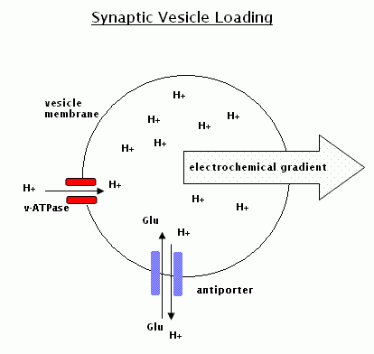
Loading of Synaptic Vesicles with Neurotransmitter
Newly synthesised synaptic vesicles are empty of neurotransmitter. They need to be filled with cargo before fusing with the plasma membrane.
Each vesicle contains 1 or 2 copies of v-ATPase within its membrane. This enzyme creates an acidic environment within the lumen of the vesicle, by using the energy of ATP hydrolysis to pump hydrogen ions (H+) inside from the cytoplasm. This creates an electrochemical gradient which provides the energy to import neurotransmitter into the vesicle via an antiporter.
E.g. For glutamate: H+ ions flow down their electrochemical gradient (out of the vesicle) and Glutamate enters via a H+/Glutamate transporter.
2. Dense Core Vesicles (DCV)
- Large vesicles with a diameter of approximately 500nm.
- They store secretory peptides e.g. hormones including catecholamines and insulin.
- Also known as secretory granules.
- Bud from Trans Golgi Network.
Formation - Dense Core Vesicles are formed in the Trans Golgi Network (TGN)
- Secretory products segregate and aggregate in the TGN.
- An immature DCV buds from the TGN.
- As the vesicle matures, its contents become more condensed. This is achieved by the formation of clathrin coated vesicles budding off the immature DCV allowing the retrieval of excess membrane and lumenal components to the golgi apparatus.
Transport to Site of Exocytosis
The mature DCV needs to be transported from the golgi to the site of exocytosis. This may be a considerable distance (e.g. in a neurone, the vesicles may need to be transported down the entire length of an axon). This movement is regulated by the cytoskeleton.
DCV's have kinesin motor proteins attached to their surface. Kinesins use energy derived from the hydrolysis of ATP to transport the vesicle along microtubules. As the DCV's reach the plasma membrane they dissociate from the microtubule and become trapped in the f-actin mesh that surrounds the cytoplasmic surface of the plasma membrane. F-actin controls docking of vesicles with the membrane.
3. Constitutive Vesicles
- Similar to dense core vesicles, constitutive vesicles bud from the TGN.
- The vesicle membrane contains proteins that contribute to the composition of the plasma membrane e.g. cell surface receptors.
- The vesicle lumen contains proteoglycans and glycoproteins that will contribute to the formation of the cells extracellular matrix once exocytosis has occured to release these products from the cell.